X-rays provide images. CT scans and MRIs provide multi-dimensional images. Endoscopic procedures provide images of what is inside the body. But there’s another type of imaging technique, and it’s one of the most intriguing. It’s called a fluoroscopy, and it essentially provides an X-ray style video of what is happening inside the body.
Consider it like a combination of an ultrasound video and something straight out of a science fiction movie.
What a Fluoroscopy Is and How It Works
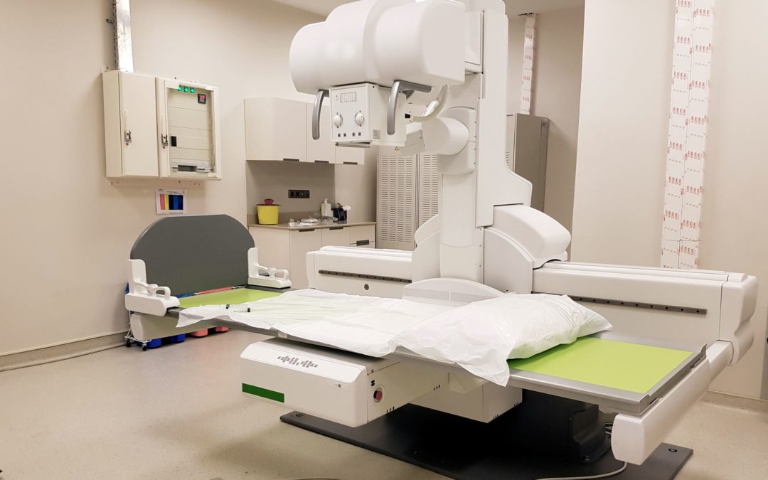
Fluoroscopy is a type of medical imaging that shows a continuous X-ray image on a monitor in incredible detail. During the procedure, an X-ray beam is passed through the body. The real-time image is transmitted to a monitor so the movement of a body part, a surgical instrument in the body, or a contrast agent can be monitored.
A fluoroscopy is like an X-ray video of what is actively happening inside your body.
A fluoroscopy can be beneficial in assisting in the diagnosis and treatment of a variety of health conditions. They are often used for:
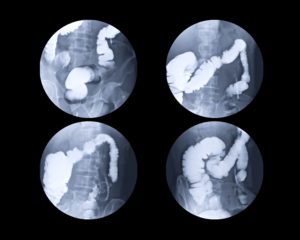
- Barium X-rays and enemas in order to view the gastrointestinal tract
- Catheter insertion and manipulation in order to direct the movement of a catheter through blood vessels, bile ducts, or the urinary system
- Placement of devices, such as stents or pacemakers, into the body
- Angiograms in order to visualize blood vessels and organs
- Orthopedic surgery in order to guide joint replacements and for the treatment of fractures
The exam itself typically takes about 20 minutes and does not generally require any preparation on the patient’s part. If a fluoroscopy is done in conjunction with other medical procedures, such as the insertion of a catheter, the duration and details depend on the requirements of the procedure.
A fluoroscopy isn’t painful and does not require any injections, anesthesia, or incisions, although the associated procedure might require these steps. However, if dye is required to enable your medical team to see clearer, this may be given to you intravenously, through an enema, or orally.
Like some other imaging techniques, the downside is exposure to radiation, although that’s typically minimal. Exposure to radiation has been associated with skin burns and some cancers, but the benefits of the imaging technique may outweigh the minimal risks in most cases.
Contact Insight Surgical Hospital for More Information
For more information about what a fluoroscopy is, how it works, how to prepare for it, or what to expect afterward, contact Insight Surgical Hospital’s health care team. We can answer any remaining questions you may have about this imaging technique or your associated surgical procedure.
Send us a message through our website or call (810) 226-0076.
Recent Comments